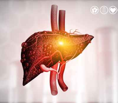
A case-study on the efficacy of pluripotent embryonic stem cell therapy in the treatment of liver failure. Read more..
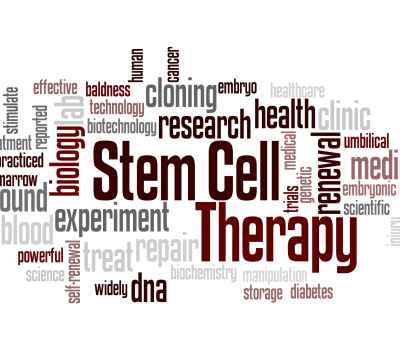
There are many clinics worldwide providing stem cell therapy, but what are the actual differences between the stem cells available? Read more..
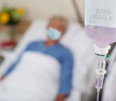
How the balancing effect of Pluripotent Stem Cells on the immune system can prevent severe damage to the lungs of those affected by COVID-19 Read more..
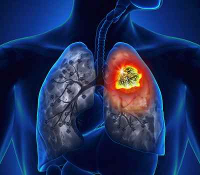
Researchers in America have discovered that vaccinating mice with embryonic stem cells prevented lung cancer in those animals that had had cancer cells transplanted into them after the vaccination or that had been exposed to cancer-causing chemicals. Read more..

A new study on mice shows that stem cells might hold the key to people maintaining strength despite their muscles’ decline that starts in their 30s. Read more..
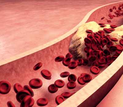
Research published in The Journal of the American Medical Association shows that a gene in Embryonic Stem Cells may play an important role in preventing the development of atherosclerotic plaques. Read more..
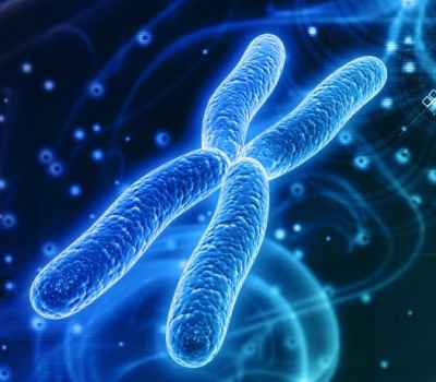
Researchers delivered a modified RNA that encodes a telomere-extending protein to cultured human cells. Cell proliferation capacity was dramatically increased, yielding large numbers of cells for study. Read more..
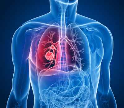
Miller School of Medicine researchers have found that the age of a donor mouse affects the ability of its mesenchymal stem cells (MSCs) to repair damage to the lungs caused by pulmonary fibrosis. Read more..
Whether you're a patient seeking stem cell therapy or a doctor seeking stem cells for your patients, please feel free to get in touch with us.
OR
Call or Text/SMS to 1-520-STEMAID